1/5

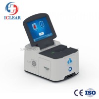
Ophthalmic Anterior Segment Analyzer Scan System
$8.00 / Piece
- FOB Price:
- Negotiable | Get Latest Price
- Order Quantity:
- 1 Set / Sets
- Supply Ability:
- 1000 Set / Sets per Month
- Port:
- shanghai
- Payment Terms:
- T/T L/C D/P D/A Credit Card PayPal Cash Escrow Other
- Delivery Detail:
- 5 days
Hot in store
-

Ce&ISO Top Grade Medical Neonatal Photot
$800.00 -

Clinical Hearing Test Audiometer Profess
$1.00 -

High Quality Home/Hospital Use Mesh Nebu
$5.00 -

Ce&ISO 8.4 Inch Medical ICU Patient Moni
$200.00 -

Heng De Faceshield with Box Transparent
$0.20 -
Whole Blood Veterinary Portable Blood Ga
$250.00 -

Endoscope Cleaning and Disinfection Devi
$1.00 -
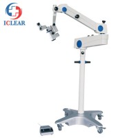
Ophthalmic Operation Microscope Stomatol
$100.00
Product Details
Product Name: Ophthalmic Anterior Segment Analyzer Scan System Model NO.: CHE-TA517 Scale: Medium Certification: CE, ISO13485 Customized: Customized Trademark: iClear Transport Package: Carton Package Specification: 505*345* 460mm 11.3Kg Origin: China HS Code: 9018500000 Product Description Ophthalmic Anterior Segment Analyzer Scan systemCHE-TA517TA517 anterior segment analyzer provides a professional solution for front and back cornea diagnosis. The device applies Scheimpflug camera which can collect 35840 data points and generates 28 cornea tomography images in high resolution. TA517 can provide a series of topography maps including cornea curvature maps, cornea thickness maps, cornea elevation maps, etc. It provides good assistance to clinicians in anterior segment diagnosis. Meanwhile, TA517 also provides chamber angle analysis, anterior chamber depth, anterior chamber volume, etc. It facilitatesclinicians in glaucoma disease diagnosis.Analytical Functions IntroductionCornea TomographyThe cornea tomography images shot by the Scheimpflug camera can give a general understanding to clinicians about patients' cornea conditions. As can be seen from above, the image displays whole cornea (from limbus to limbus). The clinician can evaluate the patient's ACD and see if the shape of the iris is normal. Meanwhile, TA517 can calculate the density of the selected area to help clinicians to see if the patients have opacity in the lens.Cornea Data Overviewbased on the images shot by Scheimpflug, TA517 can calculate a series of cornea data such as K value for front and back cornea, curvature values, etc to help clinicians has a further understanding of patient's cornea conditions.4 Maps RefractiveThe refractive maps show sagittal curvature maps for front cornea, and elevation maps for front and back cornea as well as cornea thickness map and other parameters for cornea such as steep K value, flat K value, cornea apex thickness, pupil center position and thinnest position for cornea thickness. These data are helpful in most of the cornea disease screening.Lens Fitting Analysisbased on the topography maps generated by Scansys, the system can recommend several lenses suitable for the patient's cornea and simulate the images of patient's wearing lenses with fluorescein observed by slit lamps. This will accelerate the workflow of lens fitting and save trouble for the patient to accept real fluorescein during lens fitting.Chamber Angle AnalysisTA517 can calculate a chamber angle value based on the tomography images and its exclusive AOD graph gives a trend analysis for the distance between cornea back surface to iris. It also provides cornea volume, anterior chamber volume, and anterior chamber depth calculation. These analyses are helpful to glaucoma diagnosis.Lens Density AnalysisTA517 calculates the lens density value for cross-section and longitudinal section which is helpful in cataract diagnosis.
Contact with Supplier
Recommend product
-
Stripped Soft Goose Fe
$3.00 -
plastic ball grinding
$30000.00 -
CAT piston pump 281
$4000.00 -
Droichead Zirconia Plu
$10.00 -
E.max crown, Veneer, I
Inquiry -
ReSiC Beams/plates/bur
$16.00 -
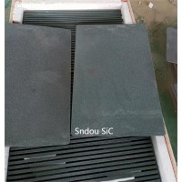
RSiC Slabs Boards Tile
$15.00 -
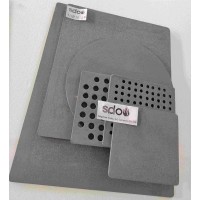
RSiC Batts as Kiln she
$15.00 -

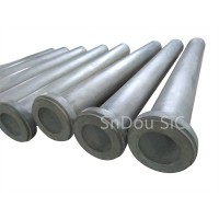
RSiC Tube by recrystal
$10.00 -
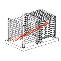
RSiC Kiln Furniture (B
$16.00 -

RSiC Burner Nozzle Fla
$18.00 -
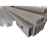
RSiC Beam Support Pill
$16.00 -
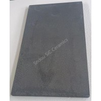
RSiC plate Slab Board
$15.00 -

NSiC Tube Pipes by Nit
Inquiry -
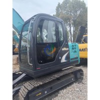
used excavator hudraul
$16600.00 -
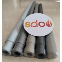
NSiC Thermocouple Prot
Inquiry -

Stalk Riser Tube for L
Inquiry -
NSiC Ceramic Heater Pr
Inquiry -

RSiC NSiC Ceramic Kiln
Inquiry -
used excavator hudraul
$11500.00
Product parameters
closure
This shop is operated by agent
- Set up shop
- Authorized by Manufacturers & Suppliers online marketplace B2B platform GongWong.com, can provide agency service
- Service Introduction
- Authorized product, Internet cloud promotion service integrating certification promotion and procurement inquiry
- Intelligent website construction
- PC terminal + mobile terminal, create a cost-effective corporate website!
closure